 The ear's 3 divisions: the outer ear, the middle ear, and the inner ear.
Anatomy Overview:
Let's learn about the structures of the human ear!
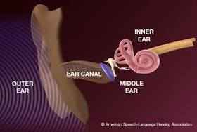
The ear consists of three sections: 1) The outer ear 2) The middle ear 3) The inner ear.
1) The pinna is the visible, cartilaginous part of the outer ear. Its job is to collect information about where sound is coming from. As you follow the pinna into the ear, you arrive in the ear canal, and at the end of this structure, you reach the eardrum, also known as the tympanic membrane.
2) The middle ear consists of the three smallest bones in the human body, which are collectively called the ossicles. Their job is to transmit pressure variations to the cochlea from sound waves that were transferred from outside of the ear to the ear drum. The stapes is an ossicle with a footplate at its end that pushes into the oval window of the cochlea.
3) The inner ear's primary structure is the cochlea. Inside this structure are the basilar membrane, tectorial membrane, and the Organ of Corti.
Each section of the system has a role in the process of hearing, all working as a unit to transmit sound to the auditory nerve.
Let's learn about the structures of the human ear!
The ear consists of three sections: 1) The outer ear 2) The middle ear 3) The inner ear.
1) The pinna is the visible, cartilaginous part of the outer ear. Its job is to collect information about where sound is coming from. As you follow the pinna into the ear, you arrive in the ear canal, and at the end of this structure, you reach the eardrum, also known as the tympanic membrane.
2) The middle ear consists of the three smallest bones in the human body, which are collectively called the ossicles. Their job is to transmit pressure variations to the cochlea from sound waves that were transferred from outside of the ear to the ear drum. The stapes is an ossicle with a footplate at its end that pushes into the oval window of the cochlea.
3) The inner ear's primary structure is the cochlea. Inside this structure are the basilar membrane, tectorial membrane, and the Organ of Corti.
Each section of the system has a role in the process of hearing, all working as a unit to transmit sound to the auditory nerve.
What are the functions and structures of the external ear?
Incoming sound waves are collected through air, and localized by the pinna. The pinna works as a filter, allowing in specific resonant frequencies between 1 and 6 kHz. Once the sound waves pass through the external ear and are lead into the ear canal, the ear canal also acts as a filter, but it is less selective; it lets through a wide variety of frequencies of the incoming sound. Once the sound waves have traveled through the ear canal, they reach the tympanic membrane (eardrum), which vibrates due to changes in pressure occurring in the ear canal by the incoming sound waves.
What are the functions and structures of the middle ear?
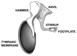 The ossicles of the middle ear, including the tympanic membrane and the footplate of the stapes.
The middle ear consists of the three smallest bones in the human body: 1) the malleus (hammer) 2) the incus (anvil) and 3) the stapes (stirrup). Collectively, these bones are called the ossicles. The job of these three small bones is to transfer sound from a mostly air filled space (the ear canal) to a fluid filled space (the inner ear). The bones of the middle ear act as a lever, increasing the force of sound wave vibrations to enter the cochlea of the inner ear. The ossicles achieve this by pushing the footplate located at the end of the stapes into a small opening on the base of the cochlea, known as the oval window. The oval window is the entryway into the cochlea of the inner ear.
What are the functions and structures of the inner ear?
The inner ear is made up of many complex structures housed within the cochlea. The most significant structures are the basilar membrane, the Organ of Corti, which sits on top of it, and hair cells (inner and outer). The basilar membrane rolled out is structured like a piano, with high notes at its stiff base, and low notes at its floppy apex. Using this piano analogy, think of piano notes as frequencies in hearing science: just like how a pressed key on the piano plays a certain pitch, each place on the basilar membrane responds to a specific frequency of the incoming sound.
Once the sound reaches the inner ear, the pressure changes in cochlear fluid allow for motion of the basilar membrane, which moves like a traveling wave from the base to the apex. Outer hair cells have little "hairs" that stick out of the top of them called stereocilia. The stereocilia are moved back and forth as the tectorial membrane brushes over them when the basilar membrane is set into its wave motion from the incoming sound source. When a wave occurs in the basilar membrane to move the outer hair cells, they lengthen and shorten (contract and expand). This lengthening and shortening of outer hair cells helps to amplify the movement of the basilar membrane at the characteristic frequency. This mechanism amplifies the basilar membrane enough, allowing us to hear at low volumes. Microscopic entities called neurotransmitters become released from the bottom of the hair cells when the tectorial membrane shears across them. The release of neurotransmitters results in action potentials occurring. These action potentials go through the auditory nerve, which has a direct connection to the bottom of the hair cells, and leads up to the brain for us to hear sounds. We hear by two ways that the ear codes sound: the rate-place code, and the temporal code.
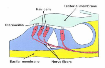
The inner ear, including prominent structures: basilar membrane, Organ of Corti, hair cells (3 rows of outer, 1 row of inner), tectorial membrane, and the auditory nerve fibers.
|
In summary:
|
